Link between OSA and the "beer gut"

General Discussion on any topic relating to CPAP and/or Sleep Apnea.
split_city
Posts: 465
Joined: Mon Apr 23, 2007 2:46 am
Location: Adelaide, Australia

Re: Link between OSA and the "beer gut"

Post by split_city » Wed Dec 23, 2009 1:25 am

Muffy wrote:
Split_City wrote:Their vertical line doesn't represent that. The difference between this figure and the Steier seated one is the that start flow doesn't coincide with end-expiration/start inspiratory effort (rise in Pdi). Thus there is iPEEP.
Are you now totally berserk? The supine example continues to show poor support for this concept and borderline accuracy at best.

Clearly, breaths 2, 3 and 4 (blue lines) do not exhibit any iPEEP characteristics, so even if breath 1 had iPEEP characteristics, it can be best qualified as an outlier:
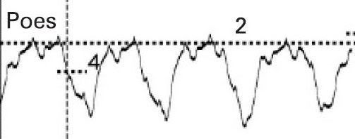
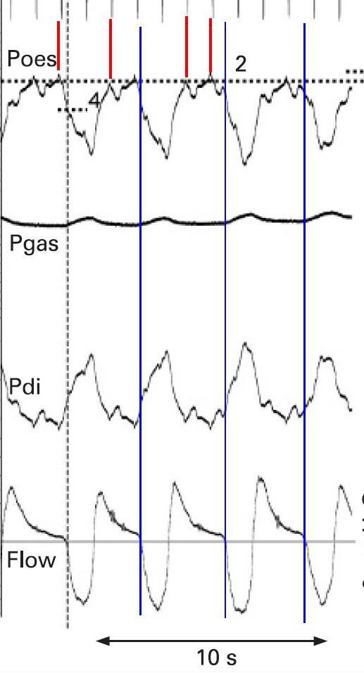
I wouldn't say breath 2 clearly had no iPEEP. Going by their definition, there still is a delay between end-expiration to start inspiratory flow. But based on what is said below, there is a question on its accuracy, which is the same with the other two breaths.
Muffy wrote:Image
Muffy wrote:One of my earlier points pertained to performing some verification of signals (biocalibrations or the like) to insure that all signals were accurate in time relative to each other. As an aside, Pankow did this:
Pankow wrote:Time synchronicity of pressure and flow measurements was assessed by placing the flow sensor, the Pao sensor, and the CMT into the tube of a bass-reflex loudspeaker. Over the range of 5–10 Hz, there was a constant time delay (Dt) of airflow in relation to pressures of 0.03 s.
but I can see no evidence of that being done in Steier (good news and bad news on that later).

However, in a review of channel synchronicity, the electrical signals seen at the top most likely represent EKG artifact. The red lines on top trace EKG down to the Pes where it can now be seen that there is a huge amount of pulse artifact contaminating the waveform, all of which needs to be subtracted from peak Pes. Consequently, that iPEEP calculation measurement would be more in the neighborhood of about 2. And given the very steep descent of Pes at that point, moving that vertical line just a tiny bit to the left (which looks quite justifiable) would give iPEEP = 0.

Muffy
Yep, all very good points. Dr. Fergal O’Donoghue, who did his PhD with us a few years back, measured iPEEP in COPD patients "Effect of CPAP on intrinsic PEEP, inspiratory effort, and lung volume in severe stable COPD. Thoax 2002." In contrast to Steier (well, it looks like it anyway), he filtered out EKG from Pes and Pdi:

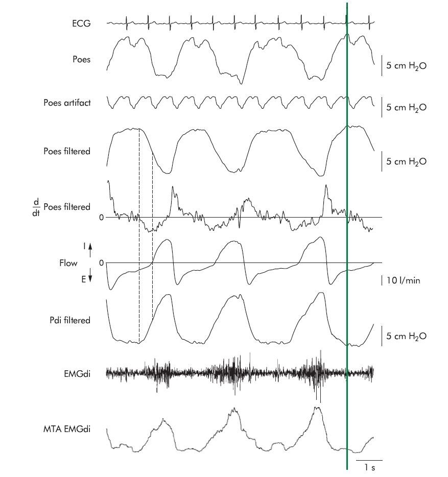
"We found that, despite optimal balloon placement, there was still
substantial cardiac artefact on Poes and Pga tracings. This
made it difficult to identify reliably the exact point of
inflection in Poes (start of inspiratory effort). Therefore, at
each CPAP pressure the mean cardiac component of the Poes
and Pga records (artefact) was calculated over a period of 2–3
minutes by ensemble averaging the recurrent pressure
fluctuations centred on the R wave of the ECG. The cardiac
artefact was found not to vary substantially within the tidal
volume range. The mean artefact was then subtracted from
the Poes and Pga traces at each cardiac cycle."

Image

Maybe that email to Steier is still worth while . Actually, since I have a litle bit of time on my hands before getting ready to sing some Xmas carols, I might just send off an email

split_city
Posts: 465
Joined: Mon Apr 23, 2007 2:46 am
Location: Adelaide, Australia

Re: Link between OSA and the "beer gut"

Post by split_city » Wed Dec 23, 2009 1:34 am

Muffy wrote:In re: the point that the Steier channels are not synchronized, it seems clear that the Pdi signal has a delay of approximately 0.3 seconds (reference the little lump following the green line), obtained by comparing it against the electrical and ballistocardiographic EKG artifact:

Image

which in a study of this nature is huge.

Unfortunately, to support my point about position of Pdi to discount the presence of iPEEP in breath 1, the shift would have needed to go to the left (flow would need the delay) but I do not see clear evidence of that surrogate biocal in the flow waveform.

Muffy
Nah, I think what they have is correct. Pdi = Pga - Pes. For simplicity, if Pga remains constant and Pes goes down, overall Pdi will go up. If Pes goes up, Pdi will go down. So it just looks like there is a delay.
Last edited by split_city on Wed Dec 23, 2009 3:18 am, edited 1 time in total.

split_city
Posts: 465
Joined: Mon Apr 23, 2007 2:46 am
Location: Adelaide, Australia

Re: Link between OSA and "Marshmallow PEEPs"

Post by split_city » Wed Dec 23, 2009 1:40 am

-SWS wrote:
Good to see you, Split. And congratulations not once but twice!
split_city wrote:my study was published in the Journal of Sleep this month...
...I have also recently submitted my PhD for review.
Thanks

User avatar
Muffy
Posts: 960
Joined: Tue Apr 21, 2009 5:28 am
Location: Schenectady, New York

Re: Link between OSA and the "beer gut"

Post by Muffy » Wed Dec 23, 2009 5:40 am

split_city wrote:Maybe that email to Steier is still worth while . Actually, since I have a litle bit of time on my hands before getting ready to sing some Xmas carols, I might just send off an email
It's not "Xmas", it's "Christmas".

Once you point out the ballistocardiographic contamination, then you can pretty much count on him going beserk, so may as well go for broke.

As noted in the O'Donoghue paper, RIP belts were used to help identify abdominal and chest activity, so ask Steier if he can produce the complete tracing of the epoch in question with RIP effort.

Since he'll probably have that study open, ask him to take away some of the flow channel filtering so that cardiac oscillations can be seen (they should be there if he fiddles enough). Preferably, get an epoch with a ill-timed beat (like a PVC). This should confirm if the flow channel is truly synched with the other waveforms.

The concept of iPEEP as being discussed here requires accepting a contradictory concept, namely, that the patient appears to be simultaneously inhaling and exhaling, and there aren't a lot of ways to do that. If RIP shows a paradoxical breath (the chest has collapsed during inspiration) in Steier breath #1 (which is what I think what really happened because iPEEP, if present in the first place, disappears in 2 breaths. What happened to that increased EELV?) then that's not iPEEP, that's a hiccup.

In looking at the O'Donoghue waveforms, there may be cardiosynchronicity in that flow channel:

Image

although those oscillations are pretty tiny, so that might be a stretch.

Muffy
________________________________

Machine: Dell Dimension 8100
Mask: 3M N-95 (during flu season)
Humidifier: Avoided, tends to make me moldy
Software: XP Pro
Additional Comments: You can't find a solution when you don't know the problem

User avatar
Muffy
Posts: 960
Joined: Tue Apr 21, 2009 5:28 am
Location: Schenectady, New York

Re: Link between OSA and the "beer gut"

Post by Muffy » Wed Dec 23, 2009 6:59 am

I just had a really funny idea!

What if ol' Barry's "expiratory instability" from days bygone was really a manifestation of the effects of iPEEP (perhaps uneven lung zone emptying). Dropping EPAP in that scenario would again be the wrong approach.

Muffy
________________________________

Machine: Dell Dimension 8100
Mask: 3M N-95 (during flu season)
Humidifier: Avoided, tends to make me moldy
Software: XP Pro
Additional Comments: You can't find a solution when you don't know the problem

split_city
Posts: 465
Joined: Mon Apr 23, 2007 2:46 am
Location: Adelaide, Australia

Re: Link between OSA and the "beer gut"

Post by split_city » Wed Dec 23, 2009 7:04 am

Talking of EPAP, I was just re-reading through another paper from the Boston group:

Sleep. 2008 Mar 1;31(3):429-32.
Effect of expiratory positive airway pressure on sleep disordered breathing.

Heinzer R, White DP, Malhotra A, Lo YL, Dover L, Stevenson KE, Jordan AS.

Sleep Medicine Division, Brigham and Women's Hospital, Harvard Medical School, Boston, MA, USA. rheinzer@post.harvard.edu

STUDY OBJECTIVES: We sought to determine the effect of expiratory positive airway pressure on end expiratory lung volume (EELV) and sleep disordered breathing in obstructive sleep apnea patients. DESIGN: Observational physiology study PARTICIPANTS: We studied 10 OSA patients during sleep wearing a facial mask. We recorded 1 hour of NREM sleep without treatment (baseline) and 1 hour with 10 cm H2O EPAP in random order, while measuring EELV and breathing pattern. RESULTS: The mean EELV change between baseline and EPAP was only 13.3 mL (range 2-25 mL). Expiratory time was significantly increased with EPAP compared to baseline 2.64 +/- 0.54 vs 2.16 +/- 0.64 sec (P = 0.002). Total respiratory time was longer with EPAP than at baseline 4.44 +/- 1.47 sec vs 3.73 +/- 0.88 sec (P = 0.3), and minute ventilation was lower with EPAP vs baseline 7.9 +/- 4.17 L/min vs 9.05 +/- 2.85 L/min (P = 0.3). For baseline (no treatment) and EPAP respectively, the mean apnea+hypopnea index (AHI) was 62.6 +/- 28.7 and 56.8 +/- 30.3 events per hour (P = 0.4). CONCLUSION: In OSA patients during sleep, the application of 10 cm H2O EPAP led to prolongation of expiratory time with only marginal increases in FRC. These findings suggest important mechanisms exist to avoid hyperinflation during sleep.

Appears there's a few issues with their P values. Probably suppose to be P = 0.03?

User avatar
Muffy
Posts: 960
Joined: Tue Apr 21, 2009 5:28 am
Location: Schenectady, New York

Re: Link between OSA and the "beer gut"

Post by Muffy » Thu Dec 24, 2009 4:43 am

Wow! That's certainly interesting, especially in light of their previous work:

American Journal of Respiratory and Critical Care Medicine. 2005 Vol 172. pp. 114-117
Lung Volume and Continuous Positive Airway Pressure Requirements in Obstructive Sleep Apnea

Raphael C. Heinzer, Michael L. Stanchina, Atul Malhotra, Robert B. Fogel, Sanjay R. Patel, Amy S. Jordan, Karen Schory and David P. White

Sleep Medicine Division, Brigham and Women's Hospital, Harvard Medical School, Boston, Massachusetts; and Rhode Island Hospital, Brown Medical School, Providence, Rhode Island rheinzer@rics.bwh.harvard.edu

Previous studies have demonstrated that lung volume during wakefulness influences upper airway size and resistance, particularly in patients with sleep apnea. We sought to determine the influence of lung volume on the level of continuous positive airway pressure (CPAP) required to prevent flow limitation during non-REM sleep in subjects with sleep apnea. Seventeen subjects (apnea–hypopnea index, 42.6 ± 6.2 [SEM]) were studied during stable non-REM sleep in a rigid head-out shell equipped with a positive/negative pressure attachment for manipulation of extrathoracic pressure. An epiglottic pressure catheter plus a mask/pneumotachometer were used to assess flow limitation. When lung volume was increased by 1,035 ± 22 ml, the CPAP level could be decreased from 11.9 ± 0.7 to 4.8 ± 0.7 cm H2O (p < 0.001) without flow limitation. The decreased CPAP at the same negative extrathoracic pressure yielded a final lung volume increase of 421 ± 36 ml above the initial value. Conversely, when lung volume was reduced by 732 ± 74 ml (n =8), the CPAP level had to be increased from 11.9 ± 0.7 to 17.1 ± 1.0 cm H2O (p < 0.001) to prevent flow limitation, with a final lung volume decrease of 567 ± 78 ml. These results demonstrate that relatively small changes in lung volume have an important effect on the upper airway in subjects with sleep apnea during non-REM sleep.

I imagine they need to do at least one more dial wingin' experiment to reconcile those differences.

Muffy

PS - I'll bet the Boston kids account for ballistocardiographic contamination in their calculations.

oxoxoxoxoxox

m.
________________________________

Machine: Dell Dimension 8100
Mask: 3M N-95 (during flu season)
Humidifier: Avoided, tends to make me moldy
Software: XP Pro
Additional Comments: You can't find a solution when you don't know the problem

split_city
Posts: 465
Joined: Mon Apr 23, 2007 2:46 am
Location: Adelaide, Australia

Re: Link between OSA and the "beer gut"

Post by split_city » Thu Dec 24, 2009 5:13 am

Which ties in nicely with their other study....

Thorax. 2006 May;61(5):435-9. Epub 2006 Feb 20.
Effect of increased lung volume on sleep disordered breathing in patients with sleep apnoea.

Heinzer RC, Stanchina ML, Malhotra A, Jordan AS, Patel SR, Lo YL, Wellman A, Schory K, Dover L, White DP.

Sleep Medicine Division, Brigham and Women's Hospital, Harvard Medical School, Boston, MA, USA. rheinzer@post.harvard.edu

BACKGROUND: Previous studies have shown that changes in lung volume influence upper airway size and resistance, particularly in patients with obstructive sleep apnoea (OSA), and that continuous positive airway pressure (CPAP) requirements decrease when the lung volume is increased. We sought to determine the effect of a constant lung volume increase on sleep disordered breathing during non-REM sleep. METHODS: Twelve subjects with OSA were studied during non-REM sleep in a rigid head-out shell equipped with a positive/negative pressure attachment for manipulation of extrathoracic pressure. The increase in lung volume due to CPAP (at a therapeutic level) was determined with four magnetometer coils placed on the chest wall and abdomen. CPAP was then stopped and the subjects were studied for 1 hour in three conditions (in random order): (1) no treatment (baseline); (2) at "CPAP lung volume", with the increased lung volume being reproduced by negative extrathoracic pressure alone (lung volume 1, LV1); and (3) 500 ml above the CPAP lung volume(lung volume 2, LV2). RESULTS: The mean (SE) apnoea/hypopnoea index (AHI) for baseline, LV1, and LV2, respectively, was 62.3 (10.2), 37.2 (5.0), and 31.2 (6.7) events per hour (p = 0.009); the 3% oxygen desaturation index was 43.0 (10.1), 16.1 (5.4), and 12.3 (5.3) events per hour (p = 0.002); and the mean oxygen saturation was 95.4 (0.3)%, 96.0 (0.2)%, 96.3 (0.3)%, respectively (p = 0.001). CONCLUSION: An increase in lung volume causes a substantial decrease in sleep disordered breathing in patients with OSA during non-REM sleep.

So basically they had three conditions in which they measured AHI:

1) baseline - no intervention (mean AHI ~62)
2) increased EELV to the same level as seen on therapeutic CPAP (mean AHI ~37)
3) increased EELV by a further 500ml (mean AHI ~31)

It can be seen that increasing EELV reduces OSA severity. However, despite EELV being at the same level induced by therapeutic CPAP, it didn't abolish events, suggesting that the the "splinting" effect of CPAP also is important. An additional 500ml increase in EELV had no apparent effect on OSA severity, which perhaps reflects a ceiling effect.

All the lung volume studies I have seen have had EELV above "normal" resting EELV and none have looked at the effect of lung volume below "normal" EELV on apnea severity. That's where my next study (effect of abdominal compression on apnea severity) is important. Stay tuned!

User avatar
Muffy
Posts: 960
Joined: Tue Apr 21, 2009 5:28 am
Location: Schenectady, New York

Merry Christmas!

Post by Muffy » Thu Dec 24, 2009 6:48 am

Image

Muffy
________________________________

Machine: Dell Dimension 8100
Mask: 3M N-95 (during flu season)
Humidifier: Avoided, tends to make me moldy
Software: XP Pro
Additional Comments: You can't find a solution when you don't know the problem

User avatar
Muffy
Posts: 960
Joined: Tue Apr 21, 2009 5:28 am
Location: Schenectady, New York

Re: Link between OSA and the "beer gut"

Post by Muffy » Fri Dec 25, 2009 6:15 am

If we can agree that the Pes and Pdi signals have not been properly synched, then it should easily follow that perhaps none of them are.

In this C&P

Image

Pes and Pdi are now synched utilizing a cardiac oscillation (red line) whose transmission should be the same in both signals. The intrathoracic portion of the pressure signal for Pes now appears a little delayed relative to Pdi (blue signal) based on nadir and peak, respectively. I moved flow over to synch with Pdi for breaths 1 and 2. This may hold for breaths 3 and 4, because the intersect (green line) may be buried by cardiac oscillation.

While flow appears dysynchronous relative to Pdi in comparing breaths 1 and 2 vs breaths 3 and 4, it cannot be moved any earlier than where I placed it because it would occur prior to Pdi in breaths 1 and 2, which would be impossible. So is the issue in all this related to breaths 3 and 4? The Pes in breath 3 is clearly the nadir. Breath 4 gets clipped by something or other, so who knows what that's from. But perhaps the real issue is that the flow of breath 3 is delayed due to airway closure.

Regardless, I think that trying to determine the presence of any iPEEP whatsoever is impossible. Even if the intrathoracic portion of the pressure signal for Pes is brought back a bit, it still would not have an iPEEP characteristic.

Muffy
________________________________

Machine: Dell Dimension 8100
Mask: 3M N-95 (during flu season)
Humidifier: Avoided, tends to make me moldy
Software: XP Pro
Additional Comments: You can't find a solution when you don't know the problem

split_city
Posts: 465
Joined: Mon Apr 23, 2007 2:46 am
Location: Adelaide, Australia

Re: Link between OSA and the "beer gut"

Post by split_city » Sat Dec 26, 2009 5:40 am

First of all, MERRY XMAS.....I mean CHRISTMAS . I hope you had a good one.
Muffy wrote:If we can agree that the Pes and Pdi signals have not been properly synched, then it should easily follow that perhaps none of them are.
Nope, I don't agree...
Muffy wrote:Pes and Pdi are now synched utilizing a cardiac oscillation (red line) whose transmission should be the same in both signals.
As I said earlier.....
split_city wrote:Nah, I think what they have is correct. Pdi = Pga - Pes. For simplicity, if Pga remains constant and Pes goes down, overall Pdi will go up. If Pes goes up, Pdi will go down. So it just looks like there is a delay.
.....I still say that their Pes and Pdi are in sync. Take the below example. I have simply derived a new channel showing Pdi. The Pes channel has a lot of ECG artefact so it's easy to identify deflections. The Pdi clearly shows the oscillations mirror that of the Pes channel.

Image
Muffy wrote: Image

The intrathoracic portion of the pressure signal for Pes now appears a little delayed relative to Pdi (blue signal) based on nadir and peak, respectively.
I guess this becomes irrelevant as they look to align in the original figure.
Muffy wrote: I moved flow over to synch with Pdi for breaths 1 and 2. This may hold for breaths 3 and 4, because the intersect (green line) may be buried by cardiac oscillation.
May or may not hold true. Depends if flow channel is really synced with pressure channels.
Muffy wrote:While flow appears dysynchronous relative to Pdi in comparing breaths 1 and 2 vs breaths 3 and 4, it cannot be moved any earlier than where I placed it because it would occur prior to Pdi in breaths 1 and 2, which would be impossible. So is the issue in all this related to breaths 3 and 4? The Pes in breath 3 is clearly the nadir. Breath 4 gets clipped by something or other, so who knows what that's from. But perhaps the real issue is that the flow of breath 3 is delayed due to airway closure.
Hhhmmm...it's all getting a little confusing! Really, we need to know whether all channels are in sync. I still say Pdi vs Pes is fine. The question is whether flow is in sync with the pressure channels. The other issue is ECG artefact. Filter that out and we might be able to tell whether iPEEP actually exists and whether it changes with posture.

I did send Steier an email. I have spoken to him in person (conference in San Fran a few years back) and also via email. I will be sure to add his thoughts once I get a reply.

User avatar
Muffy
Posts: 960
Joined: Tue Apr 21, 2009 5:28 am
Location: Schenectady, New York

Re: Link between OSA and the "beer gut"

Post by Muffy » Sat Dec 26, 2009 5:48 am

Right, since Pdi is a calculated value, in my proposal Pga would have to be moved someplace to make the formula work, and admittedly things look a little screwy when you do that (those two catheters, made in Connecticut, so you know they're high quality). However, if that's really a CO, (and I have to believe that's the case) then that must be a synch point and the other pressure measurements must have a phase delay.

Muffy
________________________________

Machine: Dell Dimension 8100
Mask: 3M N-95 (during flu season)
Humidifier: Avoided, tends to make me moldy
Software: XP Pro
Additional Comments: You can't find a solution when you don't know the problem

User avatar
Muffy
Posts: 960
Joined: Tue Apr 21, 2009 5:28 am
Location: Schenectady, New York

Re: Link between OSA and the "beer gut"

Post by Muffy » Sat Dec 26, 2009 6:16 am

On the other hand, if his calculation somehow simply results in an inversion of Pdi, that might explain a lot of things, too.

Image
Image

Muffy
________________________________

Machine: Dell Dimension 8100
Mask: 3M N-95 (during flu season)
Humidifier: Avoided, tends to make me moldy
Software: XP Pro
Additional Comments: You can't find a solution when you don't know the problem

split_city
Posts: 465
Joined: Mon Apr 23, 2007 2:46 am
Location: Adelaide, Australia

Re: Link between OSA and the "beer gut"

Post by split_city » Sat Dec 26, 2009 6:32 am

Muffy wrote:On the other hand, if his calculation somehow simply results in an inversion of Pdi, that might explain a lot of things, too.
Such as?

User avatar
Muffy
Posts: 960
Joined: Tue Apr 21, 2009 5:28 am
Location: Schenectady, New York

Re: Link between OSA and the "beer gut"

Post by Muffy » Sat Dec 26, 2009 6:55 am

Instead of Pdi = Pga - Pes, make the formula Pdi = Pes - Pga. Then you would have

Image
Image
Image

Pdi has been graphically inverted (note the label). Now the COs line up properly :

Image

Muffy
________________________________

Machine: Dell Dimension 8100
Mask: 3M N-95 (during flu season)
Humidifier: Avoided, tends to make me moldy
Software: XP Pro
Additional Comments: You can't find a solution when you don't know the problem